
Pneumothorax: Collapsed Lung Symptoms and Emergency Care
 Aug, 2 2026
Imagine you are sitting quietly, perhaps watching TV or just walking down the street, when suddenly a sharp, stabbing pain hits your chest. You take a breath, but it feels like something is blocking your lungs from expanding fully. Your heart starts racing, and you feel lightheaded. This isn't just a bad cramp or indigestion-it could be a pneumothorax, commonly known as a collapsed lung. While the term sounds terrifying, understanding what happens inside your body during this event can mean the difference between panic and effective action. It is a condition where air leaks into the space between your lung and chest wall, pushing on the lung and causing it to collapse. For some, it resolves with observation; for others, it requires immediate, life-saving intervention.
Recognizing the signs early is crucial because a simple pneumothorax can escalate into a tension pneumothorax, a deadly emergency where pressure builds up so fast that it shifts your heart and compresses your major blood vessels. In this guide, we will break down exactly what symptoms to watch for, how doctors diagnose the issue quickly, and what emergency care looks like in real-time scenarios.
What Is a Pneumothorax and Why Does It Happen?
To understand why a lung collapses, picture two layers of tissue wrapping around your lung like plastic wrap. Normally, there is a tiny amount of fluid between these layers that keeps them stuck together, allowing the lung to expand and contract smoothly with your chest wall. In a pneumothorax, air breaches one of these layers-usually the outer visceral pleura-and fills the pleural space. This air acts like a wedge, forcing the lung away from the chest wall. Without the negative pressure that normally holds the lung open, it shrinks due to its natural elasticity.
This condition doesn't happen randomly without cause, though it can seem sudden. Medical professionals classify pneumothorax into four main types based on the underlying trigger:
- Primary Spontaneous Pneumothorax: This occurs in people who do not have known lung disease. It often affects tall, thin young men (ages 20-40) due to the rupture of small air blisters called blebs at the top of the lungs. The American Thoracic Society notes this affects about 7.4 to 18 per 100,000 people annually.
- Secondary Spontaneous Pneumothorax: This happens in individuals with pre-existing lung conditions such as COPD, cystic fibrosis, or asthma. Because their lung tissue is already compromised, even a small leak can cause significant collapse and breathing difficulty.
- Traumatic Pneumothorax: Caused by external injury, such as a broken rib piercing the lung, a gunshot wound, or blunt force trauma from a car accident.
- Iatrogenic Pneumothorax: A complication resulting from medical procedures, such as lung biopsies, central line insertions, or mechanical ventilation.
Understanding the type helps determine the urgency. A primary spontaneous case might allow for a calm trip to the ER, while a traumatic or secondary case often demands rapid stabilization.
Key Symptoms: What to Look For Immediately
The hallmark symptom of a pneumothorax is acute pleuritic chest pain. Unlike the heavy, crushing pressure of a heart attack, this pain is typically sharp, stabbing, and localized to one side of the chest. It worsens significantly when you take a deep breath, cough, or move suddenly. According to clinical data from the American College of Chest Physicians, this pain often radiates to the shoulder on the same side as the collapse in over 90% of cases. If you feel a sudden stab in your right chest that shoots up to your right shoulder, pay attention.
Shortness of breath (dyspnea) is the second most common symptom, occurring in roughly 85-92% of patients. However, the severity varies wildly depending on how much of the lung has collapsed. A person with a small collapse (<15%) might only feel winded after climbing stairs, while someone with a large collapse (>30%) may struggle to breathe even while sitting still.
You might also notice physical changes in your breathing pattern. Patients often report a feeling of tightness or an inability to get a "full" breath. In more severe cases, particularly if the condition progresses to a tension pneumothorax, you may experience:
- Rapid heart rate (tachycardia), often exceeding 134 beats per minute.
- Low blood pressure (hypotension), specifically systolic pressure below 90 mmHg.
- Cyanosis, where lips or fingertips turn blue due to low oxygen levels.
- A visible deviation of the trachea (windpipe) away from the affected side, though this is a late sign and indicates critical danger.
If you have a history of lung disease and experience any new onset of chest pain or breathlessness, assume it is serious until proven otherwise. Do not wait to see if it gets better on its own.
Diagnosis: How Doctors Confirm a Collapsed Lung
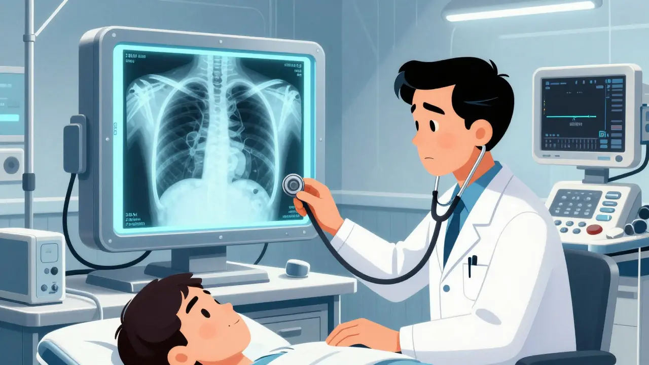
When you arrive at the emergency department, time is of the essence. The diagnostic process is designed to be rapid and accurate. The first step is usually a physical examination. A doctor will listen to your lungs with a stethoscope. In a healthy lung, you hear clear air movement. In a collapsed lung, breath sounds are decreased or absent on the affected side. They may also tap on your chest wall (percussion); a normal lung sounds dull, but a lung filled with extra air sounds hyperresonant, like knocking on a hollow drum.
Imaging confirms the diagnosis. A standard chest X-ray is the initial tool used in most hospitals. It has a sensitivity of 85-94% for detecting pneumothorax in upright patients. On the X-ray, doctors look for a visible rim of air between the lung edge and the chest wall, with no lung markings beyond that line. If the rim of air is greater than 2 cm, it generally indicates a significant collapse requiring intervention.
However, X-rays can miss small pneumothoraces, especially if the patient is lying flat (supine), which is common in trauma cases. In these situations, or when the diagnosis is unclear, a CT scan is the gold standard. A CT scan can detect as little as 50mL of air and provides a detailed view of the lung structure, helping to identify blebs or underlying diseases. Recently, point-of-care ultrasound (POCUS) has become a vital tool in emergency rooms. Using the E-FAST protocol, experienced physicians can detect the absence of "lung sliding"-the shimmering motion of the pleura during breathing-which is highly specific for pneumothorax.
| Diagnostic Method | Sensitivity | Best Use Case | Limitations |
|---|---|---|---|
| Chest X-Ray | 85-94% | Initial screening in stable patients | Misses small leaks; less accurate in supine patients |
| CT Scan | Near 100% | Unclear cases, trauma, identifying underlying causes | High radiation exposure; slower availability |
| Ultrasound (E-FAST) | 94% (experienced users) | Rapid assessment in unstable/trauma patients | Operator-dependent; requires training |
Emergency Care Protocols: From Observation to Surgery
Once diagnosed, the treatment plan depends entirely on the size of the collapse, the patient's stability, and the underlying cause. There is no one-size-fits-all approach, but guidelines from organizations like the British Thoracic Society and the American Thoracic Society provide clear pathways.
For small, primary spontaneous pneumothoraces (less than 2 cm rim) in stable patients, observation is often sufficient. The body naturally reabsorbs the air over time. Supplemental oxygen is frequently administered-not just to improve oxygen levels, but to accelerate the resorption of nitrogen from the pleural space. Studies show that high-flow oxygen can increase the rate of air resolution from 1.25% to 4.2% per hour. Patients in this category may go home with instructions to return if symptoms worsen, followed by a repeat X-ray in a few days.
If the pneumothorax is larger or the patient is symptomatic, doctors may perform needle aspiration. A large-bore needle is inserted into the chest cavity to suck out the trapped air. This procedure has an immediate success rate of about 65% for primary cases. If aspiration fails or the lung collapses again, a chest tube (tube thoracostomy) is required. A flexible tube is inserted through the chest wall and connected to a suction device to continuously remove air and fluid, allowing the lung to re-expand. This is the standard care for secondary pneumothoraces and most traumatic cases.
In the rare but catastrophic event of a tension pneumothorax, there is no time for imaging. If a patient is hypotensive, cyanotic, and struggling to breathe, doctors perform immediate needle decompression. A long needle is inserted into the second intercostal space at the midclavicular line to release the built-up pressure instantly. This is a temporary fix, followed immediately by chest tube insertion. Every minute counts here; delays increase the risk of cardiac arrest.
Long-Term Outlook: Recurrence and Prevention
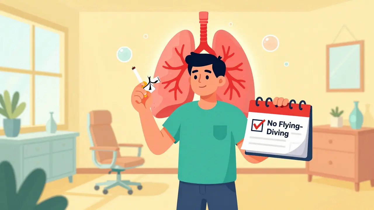
Surviving the acute episode is only half the battle. One of the biggest concerns for patients is recurrence. For primary spontaneous pneumothorax, the chance of another collapse within two years ranges from 15% to 40%. The risk skyrockets if you continue to smoke. Smoking increases the odds of recurrence by more than 20 times compared to non-smokers. Quitting smoking is the single most effective preventive measure, reducing recurrence risk by 77% within the first year.
If you experience a second collapse on the same side, doctors typically recommend definitive surgical intervention. Video-assisted thoracoscopic surgery (VATS) allows surgeons to staple off leaking blebs and perform a pleurodesis-a procedure where the lung is adhered to the chest wall using talc or mechanical abrasion to prevent future air pockets. VATS reduces the recurrence rate to just 3-5%, though it requires hospitalization and recovery time.
Lifestyle adjustments are also necessary post-discharge. The Federal Aviation Administration advises against air travel for 2-3 weeks after resolution to ensure the lung has fully healed and no residual air remains. Scuba diving is generally contraindicated indefinitely unless surgical pleurodesis has been performed, due to the pressure changes involved in diving which can trigger a recurrence.
How long does it take for a collapsed lung to heal?
Healing time varies based on the treatment method. With observation alone, small pneumothoraces may resolve in 1 to 2 weeks. If a chest tube is used, the lung typically re-expands within 24 to 72 hours, but the tube may remain in place for several days to ensure no further air leaks. Full tissue healing and return to normal activity usually take 2 to 4 weeks, depending on individual health factors.
Can a collapsed lung happen without warning?
Yes, particularly in primary spontaneous pneumothorax. It often occurs without any preceding trauma or illness. Patients may be resting or engaging in mild activity when a small air blister (bleb) ruptures. The onset is typically sudden, characterized by abrupt chest pain and shortness of breath.
Is a collapsed lung life-threatening?
It can be. While many primary pneumothoraces are manageable, a tension pneumothorax is a life-threatening emergency that can lead to cardiac arrest within minutes if not treated immediately. Secondary pneumothoraces in patients with existing lung disease also carry higher mortality rates due to reduced respiratory reserve.
What activities should I avoid after a pneumothorax?
You should avoid strenuous exercise, heavy lifting, and activities that involve significant pressure changes for at least 2-3 weeks. Specifically, avoid air travel until cleared by a doctor (usually 2-3 weeks post-resolution) and avoid scuba diving unless you have had surgical pleurodesis. Smoking cessation is critical to prevent recurrence.
How do doctors know if the lung has fully re-expanded?
Doctors confirm full re-expansion through follow-up chest X-rays. They look for the disappearance of the air rim and the return of normal lung markings up to the chest wall. In some cases, a CT scan may be used for more precise evaluation, especially if symptoms persist despite a normal X-ray.